
FileNeuron1.jpg Simple English Wikipedia, the free encyclopedia
How do you know where you are right now? Your ability to perceive your surroundings - to see, hear, and smell what's around you - depends on your nervous system. So does your ability to recognize where you are and to remember if you've been there before.
What does a neuron look like?
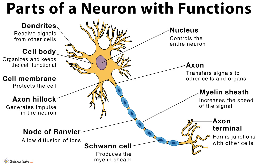
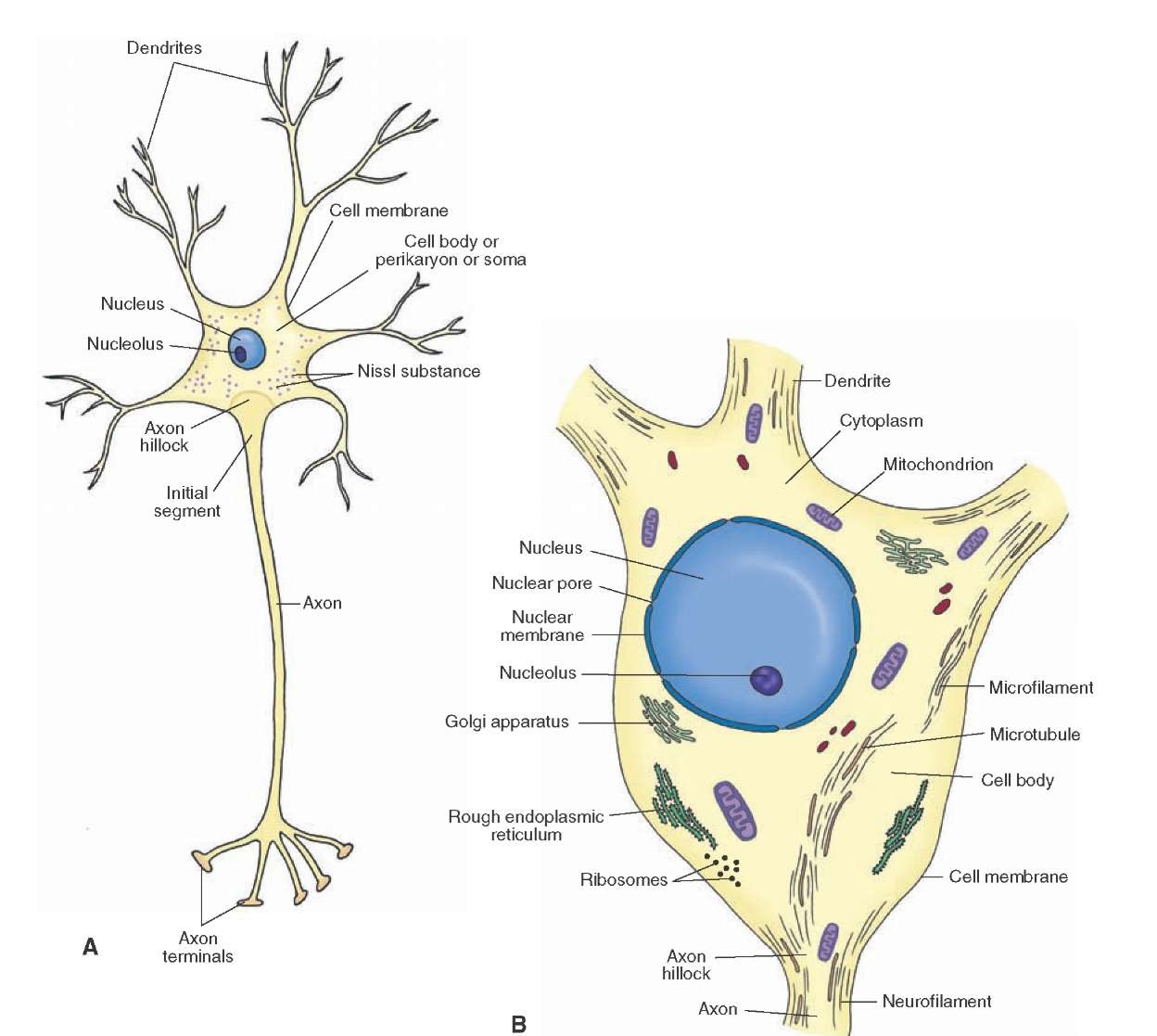
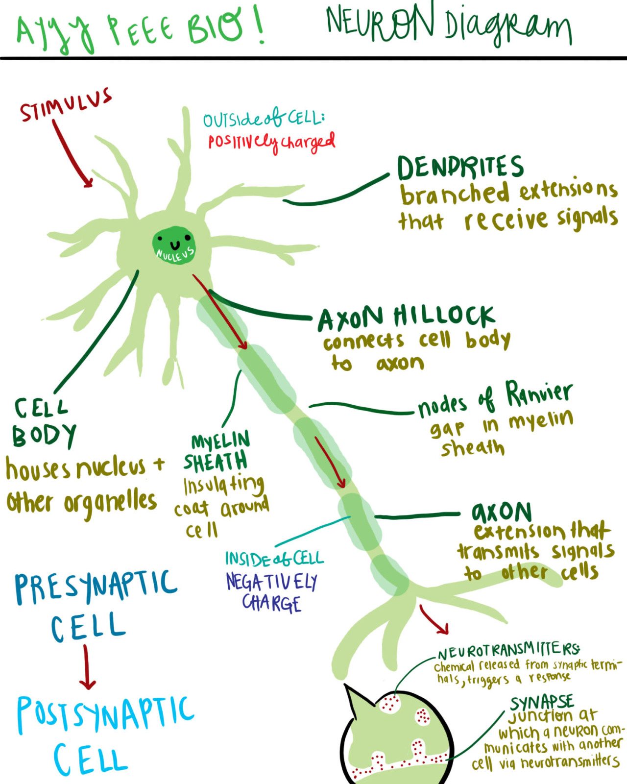
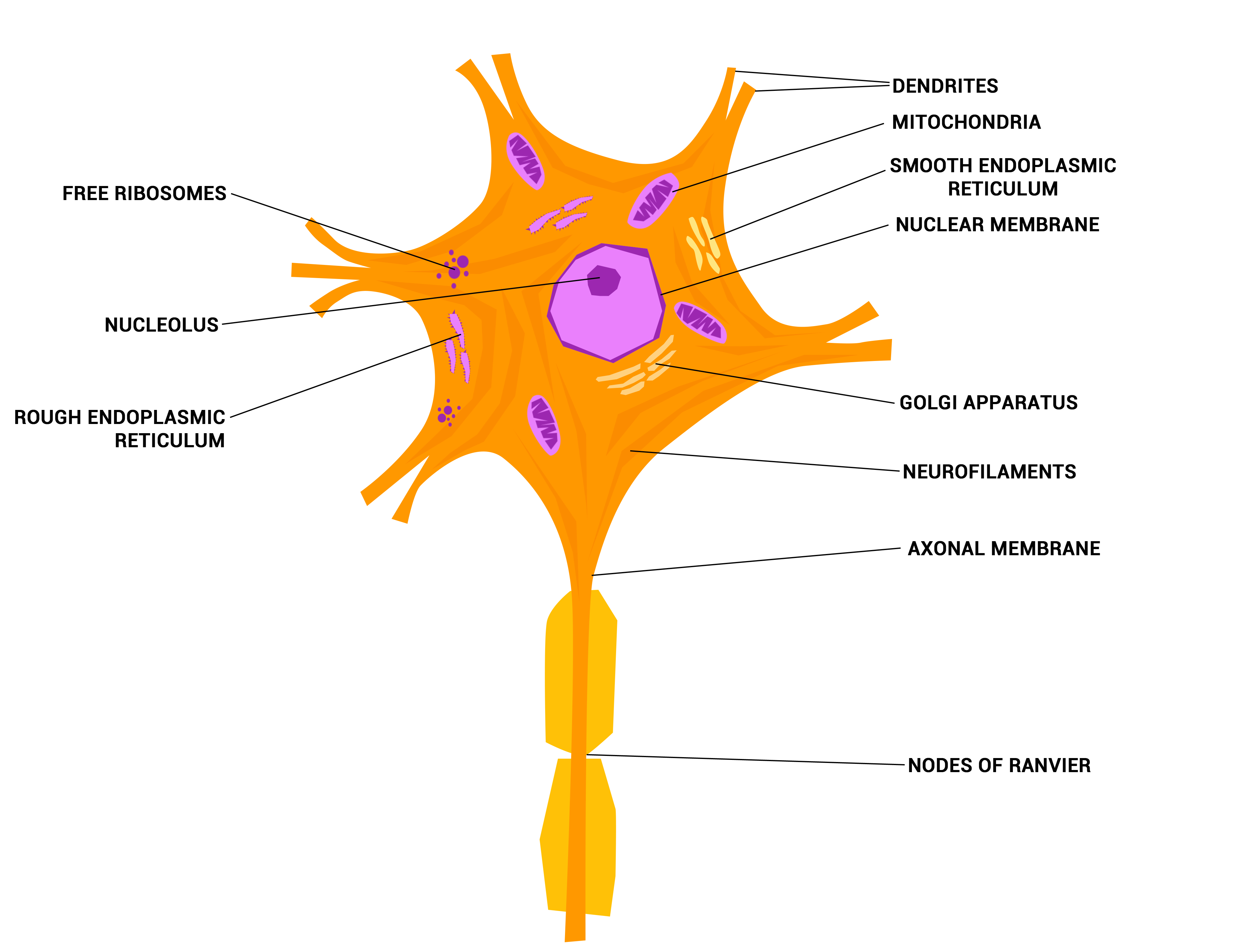
The cell body of a neuron, also known as the soma, is typically located at the center of the dendritic tree in multipolar neurons.It is spherical or polygonal in shape and relatively small, making up one-tenth of the total cell volume.. The functionality of the neuron is highly dependent on its cell body as it houses the nucleus, which contains the genetic material (DNA) of the cell as well as.
Neuron Study Guide Inspirit Learning Inc
A neuron (nerve cell) is a specialized cell that conveys electrochemical impulses throughout the body. The cytology of a neuron facilitates the transmission of either: 'top-down' information from the brain to the periphery, via efferent neurons (e.g. to permit locomotion) (efferent neurons) or,
What does a neuron look like? Biology Questions
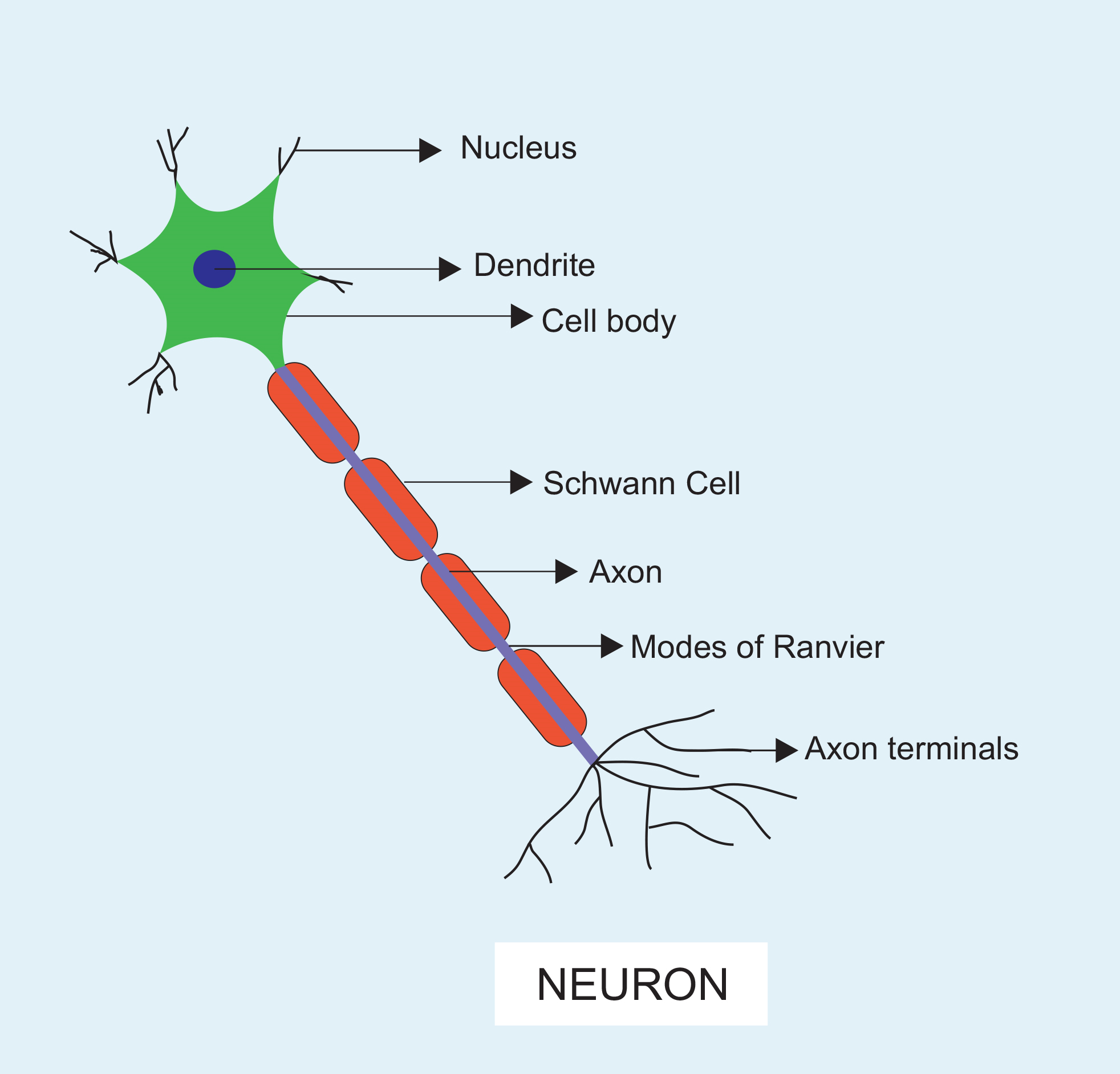
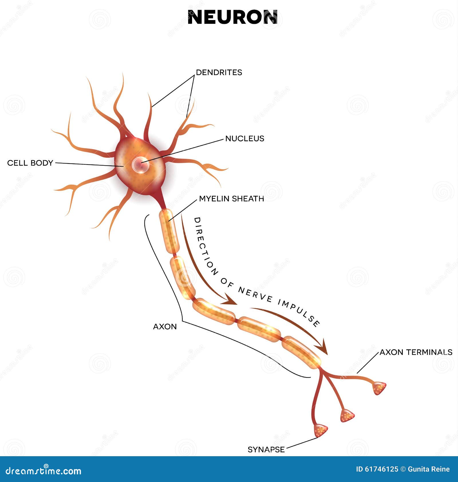
Labeled Diagram of a Neuron Definition of Neuron The building blocks of the system responsible for transmitting and processing information are called neurons. What are Neurons? Neurons are specialised cells that plays an important role in facilitating communication among different parts of our body.
Nerve Tissue SEER Training
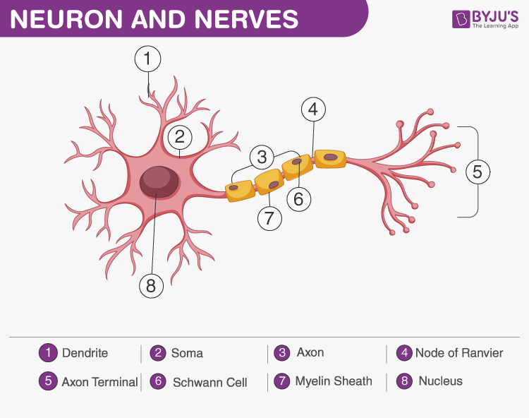
Choose the correct names for the parts of the neuron. (6) This neuron part receives messages from other neurons. (7) This neuron part sends on messages to other neurons. (8) This neuron part gives messages to muscle tissue. (9) This neuron part processes incoming messages. (10) This neuron part contains instructions for making proteins that the.
Labeled Diagram of the Neuron Stock Vector Illustration of anatomical
The nervous system is a network of neurons whose main feature is to generate, modulate and transmit information between all the different parts of the human body. This property enables many important functions of the nervous system, such as regulation of vital body functions ( heartbeat, breathing, digestion), sensation and body movements.

What Is a Neuron? Diagrams, Types, Function, and More
Neuron under Microscope with Labeled Diagram 25/04/2023 31/03/2022 by anatomylearner The structural and functional unit of the nervous system is the neuron that may easily observe under a light microscope. Neurons may vary considerably in size, shape, and other features.

The Nervous System (Structure and Function) (Nursing) Part 1
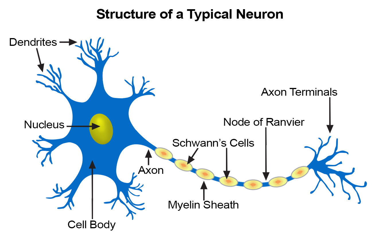
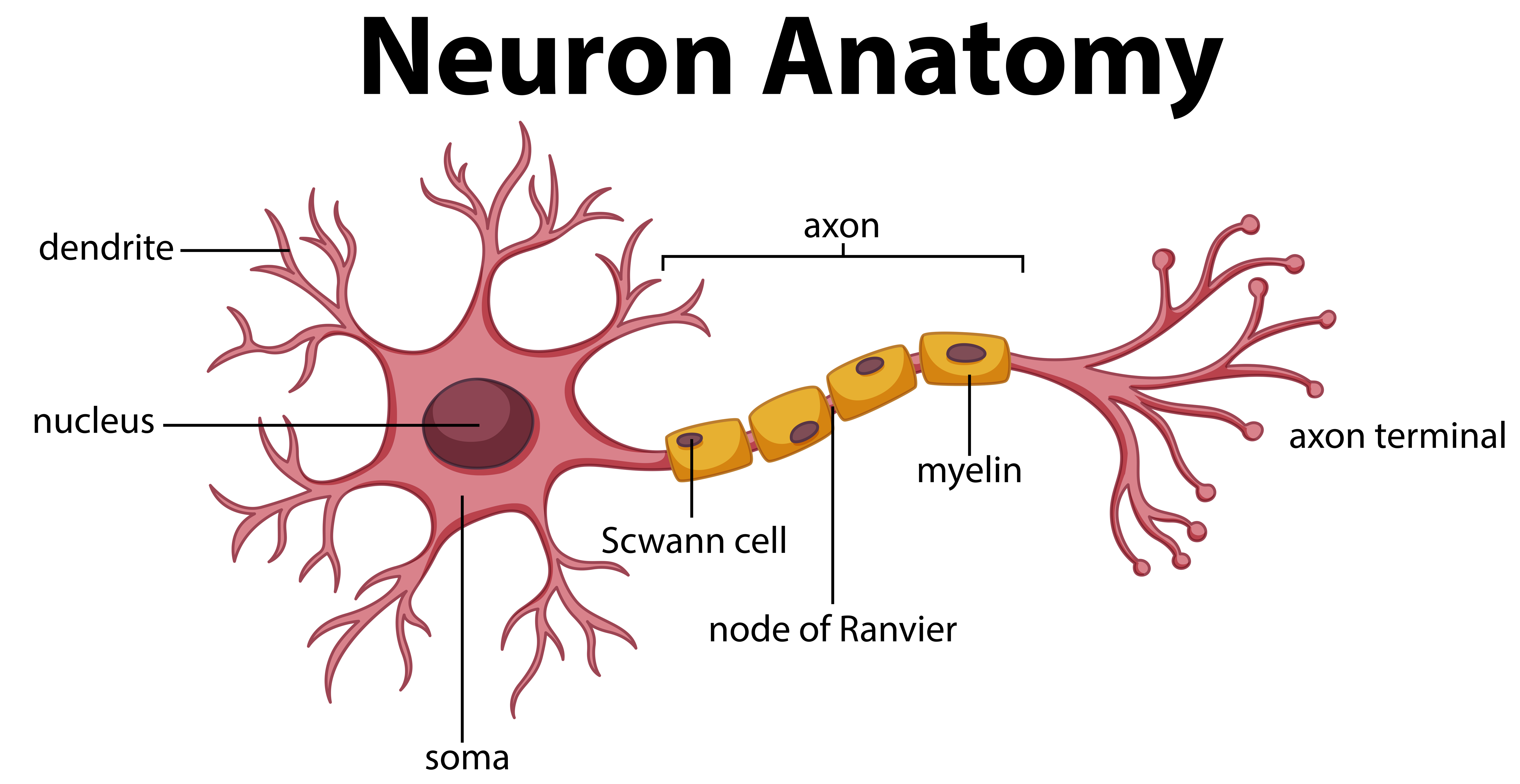
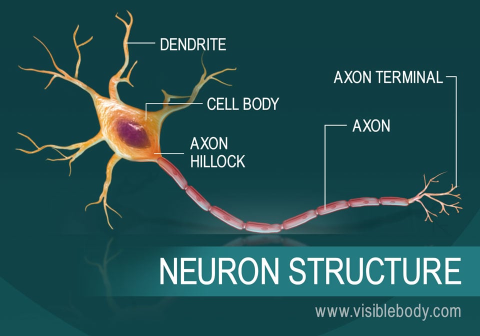
neuron, basic cell of the nervous system in vertebrates and most invertebrates from the level of the cnidarians (e.g., corals, jellyfish) upward.A typical neuron has a cell body containing a nucleus and two or more long fibres. Impulses are carried along one or more of these fibres, called dendrites, to the cell body; in higher nervous systems, only one fibre, the axon, carries the impulse.
neuron diagram labeled
Neurons or nerve cells are the basic building blocks or units of the nervous system. Nearly 86 billion neurons work co-ordinately within the nervous system to keep the body organized. They are highly specialized cells that act as information processing and transmitting units of the brain. A group of neurons forms a nerve.
Neurons
An Easy Guide to Neuron Anatomy with Diagrams Anatomy Types Function Research Takeaway Neurons, also known as nerve cells, send and receive signals from your brain. While neurons have a lot.
Histology of the Nervous System (The Neuron) Part 1
Neuron. Within a nervous system, a neuron, neurone, or nerve cell is an electrically excitable cell that fires electric signals called action potentials across a neural network. Neurons communicate with other cells via synapses, which are specialized connections that commonly use minute amounts of chemical neurotransmitters to pass the electric.
A diagram of a neuron and its functions. a study in chartreuse
3. How to Draw a Neuron Diagram To learn about the structure of the neurons, the students can use a neuron labeled diagram. The students may follow these steps to make their neuron diagram, but the process is complex: 3.1 How to Draw a Neuron Diagram from Sketch Step 1: First, the students need to draw a circle. Based on it, they need to draw a.

Neuron Diagram Straight from a Scientist
A neuron consists of neurotransmitters in the synaptic vesicles of the terminal buttons; it releases chemicals that travel across a synapse and conduct signals from one neuron to another one. Ques. Draw a labelled diagram of the neuron. (2 marks) Ans. Ques. What are receptors and what are the classification of receptors? (2 marks)
:max_bytes(150000):strip_icc()/neuron-anatomy-58530ffe3df78ce2c34a7350.jpg)
Neuron Anatomy, Nerve Impulses, and Classifications
At the synapse, the firing of an action potential in one neuron—the presynaptic, or sending, neuron—causes the transmission of a signal to another neuron—the postsynaptic, or receiving, neuron—making the postsynaptic neuron either more or less likely to fire its own action potential.

Figure 7 4 Structure Of A Typical Motor Neuron Bangmuin Image Josh
Neurons (also known as neurones and nerve cells) are electrically excitable cells in the nervous system that process and transmit information. In vertebrate animals, neurons are the core components of the brain, spinal cord and peripheral nerves. Image:Complete neuron cell diagram.svg In some countries this may not be legally possible; if so:
Neurons The crazy wires in our body. Doc Jana
Nervous tissue is composed of two types of cells, neurons and glial cells. Neurons are the primary type of cell that most anyone associates with the nervous system. They are responsible for the computation and communication that the nervous system provides. They are electrically active and release chemical signals to target cells.